November 1959 Popular Electronics
 Table
of Contents Table
of Contents
Wax nostalgic about and learn from the history of early electronics. See articles
from
Popular Electronics,
published October 1954 - April 1985. All copyrights are hereby acknowledged.
|
Optical magnification
is only useful to the point where resolution is limited by the wavelength of light
representing the object under observation. Astronomer William Dawes first provided
a means of quantification based on an ability to visually resolve closely spaced
stars. Known as the Dawes Limit, a value of 4.56/D arc seconds was empirically determined
(D is aperture of instrument in inches). A theoretical upper limit to magnification
of any optical system with perfect optics is around 2,000. As this 1959
Popular Electronics article describes, the electron microscope
removed that resolving limit by shooting a stream of electrons with radii much less
than the wavelength of visible light, and measuring its reflection. Images are necessarily
in 'false color' because we cannot perceive the real wavelength/color of the surface
revealed by the electron beam.
The Amazing Electron Microscope
 By
Morris M. Rubin By
Morris M. Rubin
Long after optical microscopes have reached their limit, the electron microscope
goes on magnifying ... up to 200,000 times.
Since the time of Anton von Leeuwenhouk - the first great microscope designer
- scientists have relied on the microscope as one of their key tools. Year after
year, as techniques for manufacturing optical glass were improved, new and better
microscopes enabled scientists to see increasingly minute objects. Then, about 1890,
it seemed that light microscopes had come to the end of the line. Beyond about 2000
times magnification, even the finest, most perfectly designed microscope showed
only a fuzzy blob.
Blocking the development of more powerful microscopes was a basic characteristic
of light itself. Similar to sound, light travels in waves of measurable length.
In the middle of the visible light spectrum, for example, the waves have a length
of about 6/250,000 of an inch. In order for light waves to differentiate between
two points of an object, they must be separated by about one-third the length of
a light wave, or about 2/250,000 of an inch. Objects smaller than about half a wavelength
cannot be magnified clearly by a light microscope, no matter how perfect its lenses.
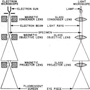
Comparison of electron microscope and ordinary light microscope.
The basic principles are the same, but the electron microscope uses coils of wire
to deflect and focus an electron beam magnetically rather than glass lenses to bend
and refract light.
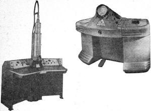
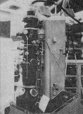
Two of the most widely used electron microscopes are shown here.
At left is the RCA EMU-3, capable of 200,000X magnification. At right is the Norelco
EM100B, which magnifies to 90,000 times.
Scientists reasoned that since the basic difficulty was caused by the relatively
long wavelengths of "ordinary" light, if it were possible to employ some type of
light which had shorter wavelengths, greater useful magnification could be achieved.
This possibility was explored, and by utilizing ultraviolet light (which has a wavelength
only one-third that of visible light), microscopes were designed which could magnify
up to 5000 times, over double the limit of visible-light microscopes.
At this point, the light microscope reached the zenith of its design capacities.
If scientists wanted more magnification, they had to find a new way of getting it.
Electrons to the Rescue.
The theory of the electron microscope was suggested in the 1920's. Experiments
showed that electrons acquired a measurable characteristic wavelength when they
were speeded up by subjecting them to high-voltage fields. The higher the voltage,
the greater was the velocity of the electrons, and the shorter became the apparent
wavelength. Further, it had been proven that electrons could be bent or refracted
by magnetic fields in much the same way light is bent and refracted by optical lenses.
Therefore, it seemed logical that light, the limiting factor in the process of
magnification, could be replaced by a stream of electrons which would have a much
shorter wavelength and so would allow greater magnification. With this important
concept under their hats, scientists began work on the design of an electron microscope.
By the late 1930's, experimental microscopes were in operation in Europe, Canada,
and the United States. And then, in 1940, RCA marketed the first commercial American
electron microscopes. These initial instruments, though crude by present-day standards,
were fantastically superior to the best light microscopes ever produced.
Whereas even an ultraviolet microscope was limited to a magnification of 5000
times, these early electron microscopes were capable of enlargements of 100,000
times. Today's models magnify more than 200,000 times - enough to see an object
one-millionth the diameter of a human hair - and by enlarging the image still further
by photographic means, useful magnifications of over one million diameters are possible!
Electrons Replace Light. Similar in principle to light microscopes,
the electron microscope uses a series of lenses to magnify the specimen in a step-by-step
process. But while a light microscope uses glass lenses to bend light rays, the
electron microscope's "lenses" are coils of wire - similar to the deflection coils
of a television set - which bend and deflect a stream of electrons.
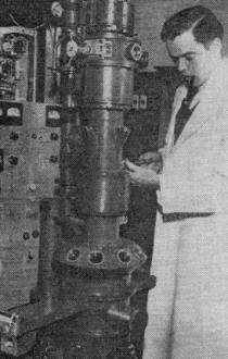
Co-inventor of the first RCA electron microscope, Dr. James Hillier,
is shown at left with RCA's Model EMB, marketed in 1940.

A 1959 Russian unit exhibited at the recent Soviet Exhibition
in New York.
Electrons emitted by the electron gun pass through the condenser lens which concentrates
the beam of electrons on the specimen. Since the specimen has been sliced so thin
as to be partially transparent, the electrons pass through it in varying numbers
depending on the density of the specimen at anyone point. Thus a pattern of varying
electron densities is produced.
Although this pattern is invisible to the eye, it could be shown by placing a
fluorescent screen below the specimen. In practice, however, the electrons pass
through the objective lens, which provides the first step of magnification. Just
before they reach the projector lens, a "spread-out" representation of the density
pattern is formed, the center area of which is then further magnified by the projector
lens.
The enlarged specimen can be viewed directly on a fluorescent screen (which looks
and works like a television screen), or the image can be photographed by special
cameras - usually built right into the electron microscope. Enlargement of the resulting
photographs allows further magnification of the specimen.
Price Tags. In addition to its optical system, an electron microscope
must have an ultra-stable high-voltage supply and a highly efficient vacuum system.
This complexity accounts for the big price tags on today's electron microscopes
- from $12,000 to $40,000, depending on the magnification desired, the make, etc.
Norelco (Philips of Holland) and RCA are the largest producers of these units.
Also active in the field are manufacturers in Germany and Japan. And the Russians
have gotten into the act, too, by producing an electron microscope which seems to
be an adaptation of a 1940 RCA model.
Limitations. Useful though the electron microscope may be, it still has its limitations.
Since the high-voltage electrons are fatal to living organisms, the electron microscope
cannot be used to view live bacteria, viruses, etc. Also, the beam of electrons
can't penetrate more than 1/25,000-of-an-inch, so the electron microscope cannot
be used to view objects that are any thicker - the wing of a fly, for example.
The solution to the latter problem has been the development of special devices
that can carve off slices of the object to be viewed which are thin enough to allow
the passage of electrons. It's easy to see how such a "slicer" works with softer
materials, but how do we go about slicing off a layer of steel 1/25,000-of-an-inch
thick? The answer to this question is surprisingly simple. A "replica" of the steel's
surface is made on a soft material - such as wax. The replica is easy to slice and,
when it is mounted on an exceedingly thin transparent membrane, it takes the place
of the original object in the microscope.
Importance. About one thousand electron microscopes are now in use in laboratories
across the nation. While they are invaluable tools in research aimed at finding
the causes of diseases, especially cancer, they are also useful in solving a wide
variety of industrial problems. For example, the wearing qualities of rubber tires
can be judged by careful examination of an electron microscope photograph, thus
eliminating the need for long and tedious road tests.
But it is in the study of cells that the electron microscope finds its most exciting
application. Cells are grown, nourished, and reproduced through a process of protein
synthesis. With the aid of electron microscopes, scientists have been able to see
these processes - which are truly the "secrets of life" - for the first time.
Man is an insatiably curious creature. One of his most efficient means of satisfying
his hunger for knowledge and understanding is the electron microscope.
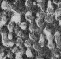
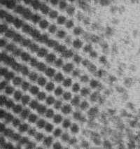
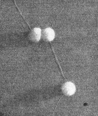
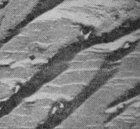
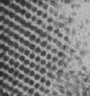
Can You Identify These Pictures?
All were taken with the aid of an electron microscope (answers are at bottom of page).
|
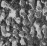
1.
(a) metal surface (b) human skin (c) leather (d) wool rug
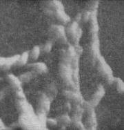
4.
(a) an ant's toe (b) polio virus (c) a sloppy knot (d) limesoap grease
|
2.
(a) wing of a fly (b) moon craters (c) protein molecule (d) cellulose
fiber
|

3.
(a) lead carbonate (b) golf balls (c) nucleic acid (d) cotton balls

5.
(a) Sahara Desert (b) silk fiber (c) quartz (d) Monday morning at
Coney Island
|
Answers
1. (a); total magnification 160,000X; courtesy of Philips Electronics, Inc.
2. (c); total magnification 425,000X; courtesy of College de France and
RCA 3. (c); total magnification 112,000X; courtesy of Dr. C. E. Hall, M.I.T.
4, (d); total magnification 68,000X; courtesy of Esso Research & Engrg.
Co. 5. (c); total magnification 14,680X; courtesy of Dow Chemical Co, and RCA
Posted June 7, 2021
(updated from original post on 5/23/2014)
|