|
May 1955 Popular Electronics
 Table of Contents Table of Contents
Wax nostalgic about and learn from the history of early electronics. See articles
from
Popular Electronics,
published October 1954 - April 1985. All copyrights are hereby acknowledged.
|
This article published in
a 1955 issue of Popular Electronics magazine is a really good primer on
the history and working principles of the electron microscope. It also explains
why such a device is needed; i.e., why an optical microscope cannot do the job when
really high levels of magnification are required. As object dimensions are spaced
at distances near to or less than the optical wavelength being observed, it becomes
impossible to resolve into separate features. Accordingly, when observing at the
upper end of the
visible light spectrum at around 400 nm,
under ideal conditions you would not be able to clearly discern two feature less
than about 800 nm apart. Current (2019)
CMOS gate thicknesses run about 5 μm, so visible light
cannot be used to image those structures. Another resolution limiting factor is
aperture size, which, depending on the wavelength causes diffraction patterns of
two objects to overlap and blend the images together. Both the
Dawe's limit
(original approximation) and
Rayleigh criterion (later, more precise) define
that resolution limit. As a long-time amateur astronomer, I am very familiar with
the aperture size limitation since a popular measure of a telescope's quality is
to determine its ability to resolve certain well-known double stars. For instance,
the North Star, aka
Polaris,
is actually a triple star system (thought to be just a double star prior to a
Hubble
Space Telescope image). Polaris A and Polaris B are separated by 18
arc seconds which according to the Dawe's limit, can be resolved with even a 50 mm
aperture if it and the eyepiece used is of high quality. The vast difference in
brightness of the two stars (magnitudes 2 and 9) complicates discernment with smaller apertures, though.
My vintage (c1969) Sears 60 mm refractor with stock eyepieces barely does the
job with my eyes which are even older than the telescope ;-(
The Electron Microscope
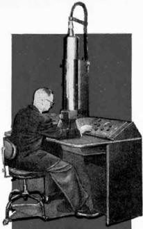 Magnifying power made possible by electronic
action provides science with a powerful weapon. Magnifying power made possible by electronic
action provides science with a powerful weapon.
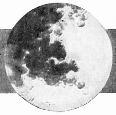
It was a vast circular plateau, high, and over two miles in diameter. It was
covered with mountains, hills, valleys, and deep galleys, and on it were ferocious
and strange beasts. The entire plateau was almost pure silver...
An imaginative description of a plateau on a remote planet as it might appear
in a science-fiction story? No! It is a description of how an ordinary dime might
appear if it could be magnified by the powerful eye of RCA's new electron microscope.
The hills, valleys, mountains, and gullies represent the markings and surface scratches
on the dime. The "strange beasts" are microscopic bacteria, germs, and virus. For
under the electron microscope, familiar objects take on strange and new appearances.
A human hair, if it could be magnified by the new microscope, would dwarf the giant
Redwoods of California. With a direct magnification of 30,000X and photographic
enlargements to 200,000X, RCA's newest commercial electron microscope has approximately
twice the magnification of earlier commercial instruments.
The electron microscope, although in use for many years, is comparatively young
as scientific instruments go, and represents one of science's most powerful weapons
in its attack on the frontier of the unknown. It has already helped to forge many
links in man's ever increasing chain of knowledge. Although the first electron microscope
was built in 1932, research and theoretical investigations which led to its development
date back many years. As far back as 1873, it was shown, theoretically, that there
was a maximum limit to the resolution that could be obtained with an optical microscope.
However, it was not until the 1920's that the need for a microscope of increased
resolving power became urgent. By this time, it was well established that a microscope
using light rays, or other types of electromagnetic radiation, could not provide
the resolution and magnification needed. Louis de Broglie, E. Schrödinger,
H. Busch, C. J. Davisson, C. J. Calbick, E. Brüche, H. Johannson, and other
scientists developed theories and practical techniques in the 20's and early 30's
which resulted in the development of practical electronic lenses.
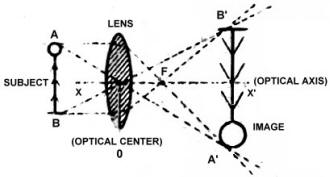
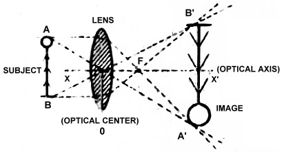
Fig. 1 - Diagram showing operation of a simple optical lens.
Converging rays of light are used to form enlarged image of a physical object. See
text for details.
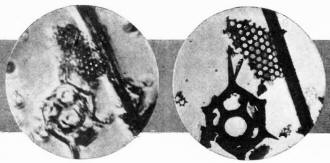
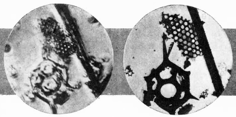
Fig. 2 - Power of RCA electron microscope is illustrated by these
two micrographs. A fossil particle was magnified 3000 times by a conventional light
microscope, and then by the electron microscope. The electron micrograph (right)
is sharp, clear, and detailed. RCA's newest unit provides magnification up to 200,000
diameters - 50 percent greater than previously possible.
After the construction of an electron microscope in 1932 by German scientists
M. Knoll and E. Ruska, work progressed at a rapid pace all over the world. In 1934,
Ruska described the construction of a new electron microscope which was built specifically
for the purpose of obtaining high resolution. Ruska's work was followed shortly
thereafter by the work of L. Barton of Brussels, and Martin, Whelpton, and Parnum
in England. Although considerable work was done from 1932 on, both in improving
the design of the basic electron microscope and in developing and refining the techniques
of using it, many of the early instruments did not provide a resolution any greater
than that obtained with existing optical microscopes.
However, scientists E. Driest and E. O. Müller, in 1935, working with Ruska's
electron microscope, and incorporating a number of changes which Ruska had suggested,
finally achieved a resolution greater than that obtained with light microscopes.
Work progressed rapidly. In 1938, B. von Borries and Ruska described an improved
electron microscope designed for practical laboratory work. Another practical electron
microscope was being designed independently of the work of Borries and Ruska by
A. Prebus and J. Hillier, two scientists in Toronto. They described their instrument
in 1939. Also in 1939, Martin designed an electron microscope for RCA and, in 1940,
RCA announced the development of a commercial electron microscope designed by Hillier
and Vance.
How It Works
In order to understand better the operation of the electron microscope, let us
first review the operation of a simple optical lens. As the reader may recall from
his high school physics, a simple converging lens may be used to form an enlarged
image of a physical object. This action is illustrated in Fig. 1. With an object
placed at AB, an enlarged image is formed at A'B'. Every ray of light entering the
lens parallel to the optical axis (XY) is bent so that it passes through the focus
point at F. Rays of light which pass through the optical center (0) of the lens
are not bent and pass straight through. At the points where the rays of light passing
through the optical center of the lens and those which enter the lens parallel to
its optical axis meet, an enlarged image of the original object is formed.
A good deal of magnification may be obtained with a simple lens of this type,
depending on the design of the lens and the relative distances between the lens,
the object, and the image. Where greater magnification is desired, a second lens
is used. The first lens forms an enlarged image, giving a certain magnification.
The second lens is located so that it magnifies the first image, giving a second,
greatly enlarged, image. The total magnification obtained, then, is the product
of the magnifications obtained with the individual lenses. For example, if the first
lens gave a magnification of 10 diameters and the second lens gave a similar magnification,
the total magnification would be 100X. Theoretically, this procedure could be carried
on indefinitely, with greater and greater magnification obtained each time another
lens is added. However, a practical limit is soon reached due to the light losses
in each lens and the loss of resolution.
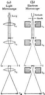
Fig. 3 - Diagram below shows comparison between a compound optical
microscope and an electron type. In the latter, an electron stream replaces conventional
light source.
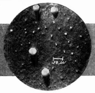
Fig. 4 - Influenza virus magnified 78,570 times. Significant
advances in medicine are made possible by unprecedented magnifications provided
by electron microscope.

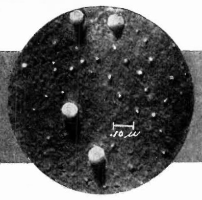
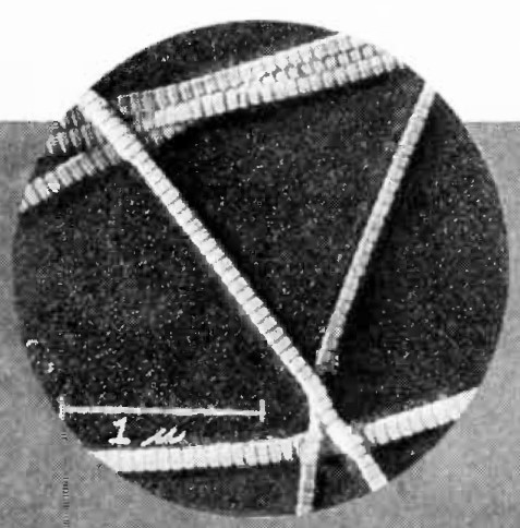
Fig. 5 - Looking like a storm cloud is this group of carbon particles
magnified 200,000 times under an electron microscope. Studies like this provide
valuable data to industry.
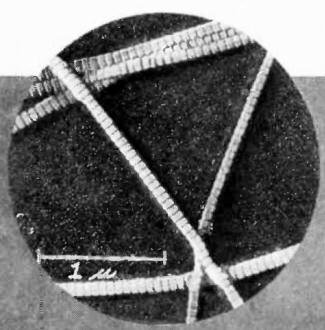
Fig. 6 - Human collagen is magnified 34,285 times by RCA electron
microscope to provide scientists with close-up study of essential ingredient of
bone's organic substance.
There are two factors which are important when dealing with microscopes, magnification
and resolution. The magnification is, of course, the ratio of the sizes of the image
and the original object. The resolution is essentially the ability of the microscope
to distinguish between two small points close together. The two do not necessarily
go together. It is possible to have a microscope of high magnification but with
poor resolution, and small details, although greatly magnified, may tend to blur
together. See Fig. 2.
Due to the nature of light and lenses, there is a practical limit to the amount
of resolution that can be obtained with optical microscopes. The observation of
extremely small objects is limited by the wavelength of the light used, with the
best microscopes distinguishing objects whose size is about half that of the wavelength
of the light used. The practical limit is reached when light of extremely short
wavelength, such as deep blue or ultraviolet, is employed.
The smallest particle that can be seen, even with the finest optical microscopes,
must have linear dimensions of at least 1/125,000 of an inch, and the greatest useful
magnification is, therefore, only about 2000X. Under special circumstances, optical
magnifications up to 5000X may be used. Unfortunately, scientists find it necessary
to observe viruses and other tiny objects which may have dimensions of only 1/1,000,000
of an inch. Trying to see these extremely small objects with light is almost like
trying to observe a mouse by throwing a basket full of tennis balls at it and noting
which ones are bounced back. It was this impasse that led to the development of
the electron microscope ... for electrons are much smaller, physically, than the
wavelength of even ultraviolet light, and thus may be used to observe extremely
small objects. Theoretically, an electron microscope may be used to observe molecules
and, perhaps, individual atoms!
A combination of lenses to obtain increased magnification is known as a compound
microscope. The first lens in the system is generally called the objective lens
and the last one may be called either the projecting lens if the image is thrown
on a screen or photographic plate, or an eyepiece lens if the image is viewed directly
with the eye. A direct comparison of a compound optical microscope and an electron
microscope is given in Figs. 3A and 3B, respectively.
In the light microscope, shown in Fig. 3A, a lamp acts as a source of light.
The light rays are concentrated and formed in parallel beams by the condensing lens
L1. These light beams strike the subject object S, outlining it. In addition,
greater or lesser amounts of light may pass through the subject, depending on its
relative transparency. The majority of light microscopes depend on light passing
through the object to be viewed. An enlarged image of the subject is formed by the
objective lens L2 and appears at I. This image, or, actually, a portion
of the entire image, is enlarged by the eyepiece or projecting lens L3,
with the second image appearing at I2. The final image is a "light shadow"
of the subject.
Point-by-point, the electron microscope works in a very similar fashion. Instead
of the lamp which served as a light source, a combination of a cathode and an anode
serves as an electron source (such a combination is frequently called an electron
"gun "), for in the electron microscope light rays are replaced by streams of electrons.
Electrons are "boiled" out of the heated cathode and are accelerated forward by
the positively charged anode. They are then concentrated and focused into a sharp
beam by the condensing magnetic lens L1. The action of a magnetic lens
is to bend and to focus the stream of electrons in much the same way that a glass
lens bends and focuses a light beam. A magnetic lens is made up of a soft iron shell
enclosing a coil of copper wire. A narrow ring-shaped gap is made on the inside
of the iron shield and this gap, together with the amount of current flowing through
the coil, determines the characteristics of the magnetic field produced and hence
the "optical" characteristics of the lens.
The focused electron beam strikes the subject object S. As in the case of the
light microscope, the electrons must pass through the object and thus the subject
must be relatively transparent to the stream of electrons. The objective lens L2
forms an enlarged image of the subject at I. A portion of this image is magnified
by the projecting lens L3 which forms the final image at I2.
The final image is an "electron shadow" of the subject.
In an optical microscope, the light is visible, of course, and it is only necessary
to allow the light to strike a plain white reflecting surface for the image to be
seen. In the electron microscope, on the other hand, the electrons are not visible,
so the electrons are allowed to strike a fluorescent screen to form the final image.
The screen used is similar in composition to the fluorescent screen used on the
face of a cathode-ray tube in a television receiver. In either the optical or the
electron microscope, sensitive photographic film or plates may be substituted for
the viewing screens. The picture obtained may then be enlarged photographically
to give even greater magnification. This is quite feasible in the case of the electron
microscope, since resolution is good. Considerable photographic enlargement is possible.
In the light microscope, however, the poor resolution obtained limits the amount
of photographic enlargement that may be used before the image becomes too blurred
to be useful.
In the future, we may expect to see even more extensive use of the electron microscope
than we have in the past. It may well be that the electron microscope will play
an important role in defeating the remaining disease enemies of mankind.
Posted September 10, 2019
|